
Short answer
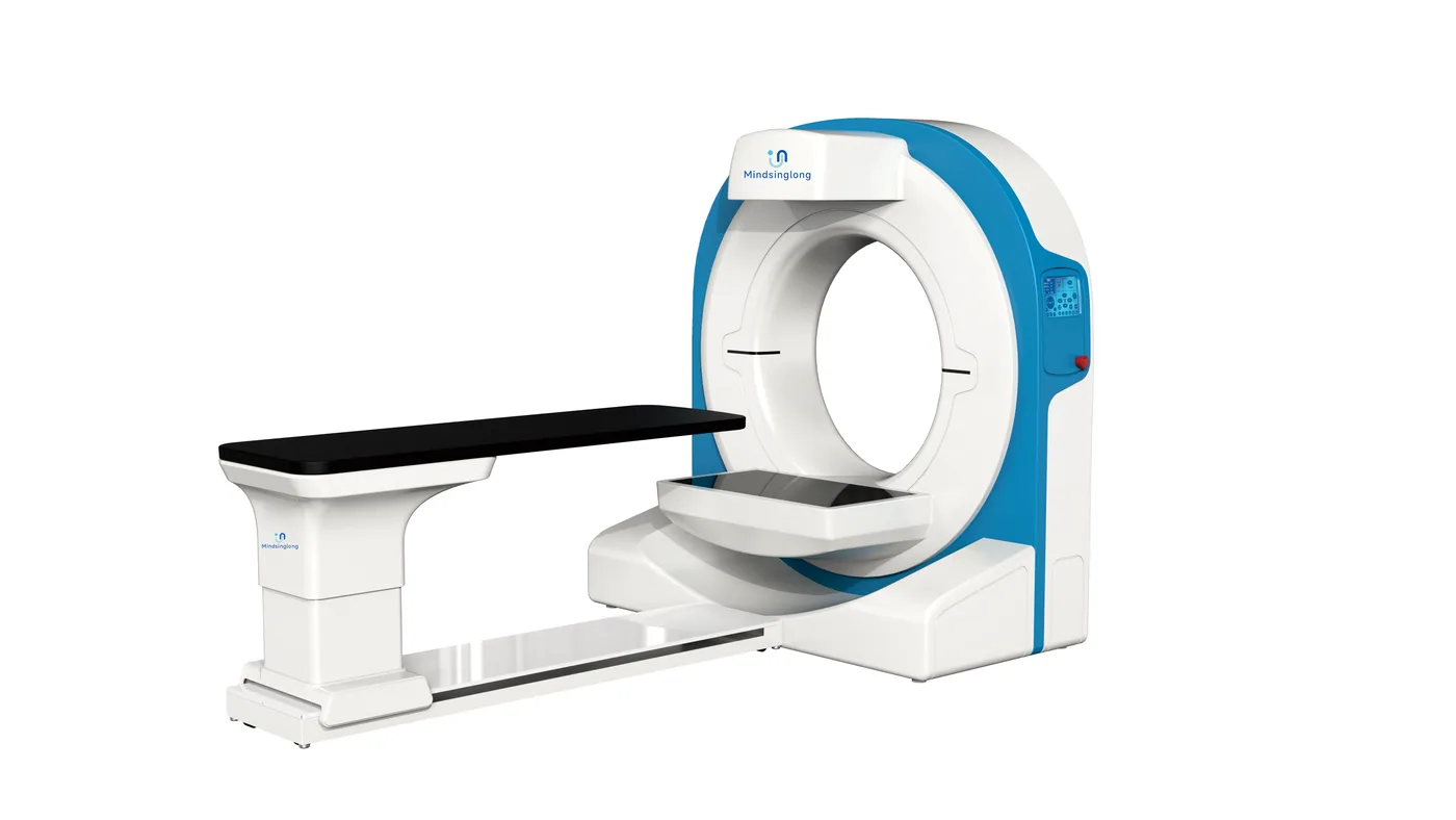
A veterinary CT scanner purchase should begin with clinical questions and patient geometry, not slice count or purchase price. Define the species, weight range, target anatomy, expected case volume and reporting pathway. Then compare scanner geometry, acquisition and reconstruction, motion sensitivity, anesthesia integration, radiation protection, room infrastructure, service coverage and complete ownership cost. Conventional fan-beam CT and cone-beam CT can have different strengths; neither label guarantees suitability for every soft-tissue, orthopedic, dental or exotic-animal application.
Editorial note: This independent procurement guide was prepared from Mevidon's supplied product materials and the primary professional or government sources listed below. It contains no paid rankings and does not replace clinical, engineering or regulatory advice.
Quick comparison
| Planning area | Question to resolve | Evidence to request |
|---|---|---|
| Clinical scope | Which anatomy and decisions? | Representative studies and protocol list |
| Patient fit | Weight, width, length and positioning? | Aperture, table and load specifications |
| Image workflow | Who acquires, reconstructs and reports? | Demonstration, DICOM and workstation scope |
| Facility | Room, power, cooling and shielding? | Final pre-installation document |
| Uptime | Who responds when the system stops? | Warranty, parts and service-level terms |
Which clinical applications justify the project?
List the referral and in-house questions the scanner is expected to answer: for example selected cranial, dental, orthopedic, thoracic, abdominal or surgical-planning applications. Avoid broad claims such as whole-body or all species without protocol-level evidence. Estimate monthly cases, current referral leakage, anticipated report turnaround and how results change management.
Create an acceptance set using anonymized representative cases or phantoms. The intended radiologist and operators should review image quality, artifacts, reconstruction tools and reporting workflow before the final commercial decision.
How should patient size and scanner geometry be specified?
Weight capacity alone is not enough. Record maximum patient width, length, posture, target anatomy and the space needed for supports, tubes and monitoring lines. Compare gantry aperture, field of view, table travel, table load, positioning aids and access to the airway. Large dogs and small exotics create different geometry and motion challenges.
Ask the supplier to confirm the usable scan region for your required positions, not only the nominal aperture. Review emergency removal and patient transfer with the clinical team before installation.
Cone-beam CT or conventional CT?
Cone-beam CT uses a cone-shaped X-ray beam and flat-panel detector to acquire a volume, while conventional CT typically uses fan-beam geometry with detector rows. Performance depends on the complete design, protocols and reconstruction. CBCT can be attractive for selected dental, cranial and orthopedic workflows; conventional CT may be preferred for broader cross-sectional applications. These are tendencies, not purchase rules.
Request same-task evidence for the anatomy, patient size and clinical question. Compare soft-tissue contrast, spatial resolution, motion and metal artifacts, scan field, acquisition time and dose information. Do not infer performance from detector or slice terminology alone.
What acquisition, reconstruction and data features matter?
Review acquisition time, protocol selection, slice or voxel options, multiplanar reconstruction, 3D tools, metal-artifact handling, measurement functions and export formats. Confirm what is automatic, what requires manual reconstruction and which packages cost extra. Evaluate the diagnostic workstation separately from the scanner console.
DICOM storage, worklist and routing should be tested with the clinic's PACS or teleradiology provider. Estimate study size, daily storage, backup and network upload time. Cybersecurity responsibilities, user accounts, software updates and remote-service access belong in the contract.
How should anesthesia and patient monitoring be integrated?
Motion can degrade CT data, so the veterinary team must define immobilization, sedation or anesthesia requirements for each protocol. The project may need an anesthesia machine, compatible breathing circuits, patient monitor, oxygen source, infusion support and a recovery area. Position equipment so staff can observe and access the patient while respecting radiation-protection controls.
The scanner supplier should provide interface and room constraints, while qualified veterinary staff establish clinical protocols. Conduct a simulated patient journey from preparation through positioning, scanning, emergency removal and recovery before the first clinical case.
What radiation and facility planning is required?
CT uses ionizing radiation. Engage the local regulator and qualified radiation-protection expert early. The IAEA veterinary safety report addresses occupational and public exposure, controlled areas, equipment and safe operation. Local rules determine shielding design, registration, personnel qualifications, dosimetry, warning systems, quality assurance and recordkeeping.
Obtain final dimensions, weight, heat load, power quality, grounding, cooling, access route, control-room layout, network, fire safety and service-clearance requirements for the exact configuration. Freeze the equipment model before approving construction drawings.
How do you protect uptime and serviceability?
CT downtime affects referrals, patient scheduling and revenue. Identify who performs installation, acceptance testing, preventive maintenance and corrective service. Ask for remote-response hours, on-site coverage, engineer location, parts location, escalation path and exclusions. Clarify responsibilities for the X-ray tube, detector, computer, software, UPS and third-party accessories.
A realistic ownership model includes planned maintenance, replacement risk, software support, service travel, quality-control testing and lost capacity during downtime. Obtain references only when they can be verified and are relevant to the same market and scanner class.
What should be included in the final quotation?
The quotation should list the gantry, patient table, console, reconstruction and diagnostic workstations, software packages, positioning aids, contrast accessories if required, anesthesia and monitoring items, UPS, networking, installation, testing, training, documentation, warranty, maintenance and delivery terms. It should also state what the buyer or local contractor provides.
Mevidon can help compare configurations and coordinate international supply. Final clinical suitability, radiation approval, compliance documents and facility design must be confirmed for the selected model and destination market before purchase.
Copy this buyer checklist
- Which anatomy, patient sizes and clinical decisions define acceptance?
- Does the project require CBCT, conventional CT or a modality comparison?
- How will motion, anesthesia, airway access and recovery be managed?
- Who will interpret studies and what DICOM workflow is required?
- Who approves radiation protection and the final room design?
- What electrical, cooling, network and access constraints apply?
- What warranty, parts, response time and maintenance costs are documented?
Buyer FAQs
What is the best veterinary CT scanner?
There is no universal best scanner. The right system must fit the clinic's patients, anatomy, clinical questions, room, workflow, reporting capacity, service coverage and budget.
What is the difference between veterinary CBCT and CT?
They use different acquisition geometries and may differ in field of view, contrast, spatial resolution, motion behavior and workflow. Buyers should compare representative studies and protocols instead of relying on the label.
How much space does a veterinary CT scanner need?
Space depends on the exact scanner, control area, shielding, service clearances, patient preparation and anesthesia workflow. Use the final vendor pre-installation document and a locally approved room plan.
Can CT images be sent for remote interpretation?
Often yes when DICOM export, adequate connectivity and a compatible teleradiology workflow are available. These interfaces and expected study upload times should be tested before acceptance.
Sources and further reading
Primary sources were accessed and checked on July 19, 2026.
- FDA: Medical X-ray ImagingDefines radiography and CT, and explains justification, optimization and ALARA principles.
- FDA: Computed Tomography (CT)Explains cross-sectional CT imaging, benefits, radiation considerations and facility responsibilities.
- IAEA Safety Reports Series No. 104Veterinary-specific guidance for worker and public radiation protection, facility planning and safe operation.
Important procurement note
This guide is educational and does not replace clinical, engineering, radiation-safety, occupational-safety, regulatory or facility advice. Product capability and compliance depend on the final model and market. Confirm the signed specification, included accessories, local requirements, installation scope and service plan before purchase.