
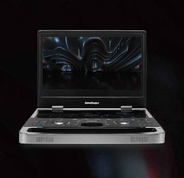
Veterinary ultrasound is one of the most versatile imaging tools in animal healthcare. It provides real-time views of soft tissue and motion without ionizing radiation, supporting routine examinations, urgent decisions, reproductive work and specialist imaging across companion animals, livestock and equine practice.
What is a veterinary ultrasound machine?
A veterinary ultrasound machine sends high-frequency sound waves into the body through a transducer. Reflections from tissues are processed into an image on the display. Because the image updates in real time, the operator can observe organ movement, guide selected procedures and assess blood flow when Doppler modes are available.
A complete system is more than the main console. The probe set, imaging modes, battery, software, measurements, storage, reporting, cleaning workflow and service plan all affect clinical usefulness. The right system therefore depends on what a veterinary team needs to examine, not on one headline specification.
Common clinical applications
Abdominal and soft-tissue imaging
Ultrasound can support evaluation of the liver, gallbladder, kidneys, bladder, spleen, gastrointestinal tract and other soft tissues. Image quality depends on probe frequency, patient size, acoustic access and operator technique.
Reproduction and pregnancy
Veterinary teams use ultrasound for pregnancy detection and reproductive assessment in companion animals, cattle, sheep, goats, pigs and horses. A portable system can be particularly useful for field examinations and herd work.
Cardiac and vascular assessment
Cardiac examinations may require phased-array transducers, M-mode, color Doppler and spectral Doppler. The necessary configuration changes with patient size and the level of cardiac measurement expected.
Musculoskeletal and point-of-care use
High-frequency linear probes can support superficial structures, tendons, ligaments and selected joints. Point-of-care ultrasound can help rapid triage and focused assessment when integrated into a defined clinical protocol.
Types of veterinary ultrasound systems
| System type | Typical advantage | Questions to confirm |
|---|---|---|
| Handheld or wireless | Maximum mobility and rapid deployment | Compatible display, battery, probe geometry, cleaning and data storage |
| Portable laptop-style | Balance of mobility, controls and imaging capability | Weight, active probe ports, display, Doppler modes and cart options |
| Cart-based | Dedicated ergonomics, larger display and advanced configuration potential | Room space, electrical needs, probes, software packages and service |
| Black-and-white | Focused routine or reproductive imaging at a lower entry level | Clinical limits, measurements, image storage and upgrade path |
| Color Doppler | Blood-flow visualization and broader abdominal, vascular or cardiac use | Color, power, PW/CW Doppler, sensitivity and probe support |
How to choose the right veterinary ultrasound machine
- Define the animal range. List the smallest and largest patients and whether the work is small-animal, equine, livestock or mixed practice.
- List the examinations. Separate abdominal, reproductive, cardiac, vascular, musculoskeletal and interventional needs.
- Choose the probe set. Confirm transducer type, frequency range, footprint and the number of active ports required.
- Match the imaging modes. Decide whether B-mode is sufficient or whether M-mode, color, power, PW, CW, 3D or 4D capabilities are justified.
- Review the workflow. Consider portability, display, controls, battery time, storage, reporting, DICOM, cleaning and the examination environment.
- Compare the total scope. Include probes, cart, printer, software, measurements, warranty, training, shipping, documentation and after-sales support.
For a procurement-focused checklist, read Mevidon’s veterinary ultrasound machine buying guide. You can also compare the portable and cart-based formats.
From requirement to confirmed configuration
Mevidon begins with a written requirement brief covering species, clinical applications, workload, facility conditions, destination market and budget. A useful quotation should identify the main unit, included probes, optional software, accessories, documentation, training, warranty, lead time and exclusions. Product certification and regulatory suitability must be confirmed for the selected model and destination rather than assumed from a general category statement.
Buyers can review the portable veterinary ultrasound machine and veterinary color Doppler ultrasound machine pages for current product evidence and configuration examples.
Frequently asked questions
What is a veterinary ultrasound machine?
It is a diagnostic imaging system that uses high-frequency sound waves to create real-time images of internal organs and soft tissues in animals.
Is veterinary ultrasound safe?
Diagnostic ultrasound does not use ionizing radiation. Examinations should still be performed by trained veterinary professionals using appropriate settings and protocols.
What is the difference between black-and-white and color Doppler ultrasound?
Black-and-white imaging shows anatomy and tissue structure. Color and spectral Doppler add information about blood-flow direction and velocity for vascular and cardiac assessment.
Should a clinic choose a portable or cart-based system?
Portable systems favor mobility and flexible room use. Cart-based systems can offer larger displays, dedicated controls and more advanced configurations. The better choice depends on exams, workload and budget.
Which probes are most useful in veterinary practice?
Convex or micro-convex probes are common for abdominal and reproductive work, linear probes for superficial structures, and phased-array probes for cardiac examinations.
Technical reference
The U.S. Food and Drug Administration explains that diagnostic ultrasound creates images using high-frequency sound waves and does not use ionizing radiation. See FDA: Ultrasound Imaging. Clinical interpretation and examination protocols remain the responsibility of trained veterinary professionals.
Need help matching a veterinary ultrasound configuration?
Send Mevidon your animal range, examinations, preferred format, destination and budget for a structured comparison.
Discuss your requirement →